Separation of Histone H1.5

Get In Touch
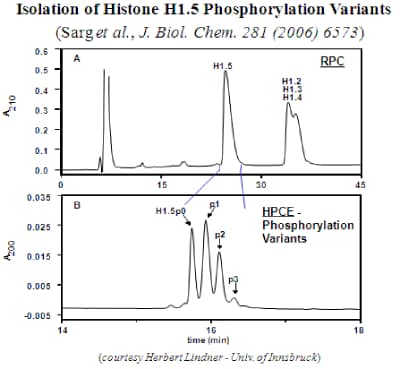
HPLC is superior to electrophoresis for separation of protein variants. The figure below shows the isolation of Histone H1.5 and the separation of phosphorylation variants by HPCE.
Application Name : Isolation Of Histone H1.5 phosphorylation variants
Column Name : PolyCAT A™ Columns
Analytes : Histone H1.5

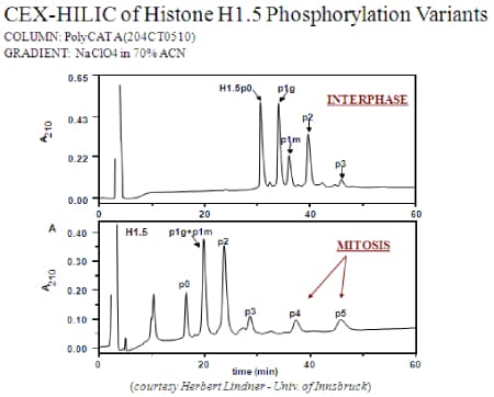
When the same mixture is resolved by cation-exchange in the HILIC mode [BELOW], the resulting peaks are more than twice as sharp. Furthermore, the column is able to distinguish between positional variants, each bearing a single phosphate group but on different Ser- residues. When the cell is in mitosis, more highly phosphorylated forms appear that are absent in interphase. Again, these forms are required for successful entry of the cell into mitosis. The schematic below shows the sequence of phosphorylation. The variants with 4 or 5 phosphates represent the first observation of a histone phosphorylated at a Thr- residue. Again, these low-abundance forms could only be identified by first separating them prior to digestion from more abundant variants.
Application Name : CEX-HILIC Of Histone H1.5 Phosphorylation Variants
Column Name : PolyCAT A™ Columns
Analytes : Histone H1.5
Get Your Quote or Call: 040-29881474
We focus on supporting laboratory workflows & optimizing lab-wide operations
